Publications
3,325
Number of publications (2024)

The Faculty of Medicine and LMU University Hospital are among the strongest reseach centers of university medicine in Europe. Scientists develop new diagnostic methods and treatment options and train the next generation of scientists. Patients benefit from cutting edge medicine in clinical trials. Patients benefit directly from the findings of cutting-edge medicine in clinical studies.

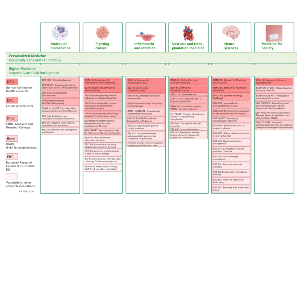
As shared facilities, core facilities offer scientists access to large-scale research equipment, modern technologies and scientific services.
Read more
Scientists use the first-class infrastructure of the Munich Science Network and cross-location infrastructures.
Read more
46 clinical departments, divisions and institutes at LMU University Hospital conducted a total of 2,322 clinical studies in 2022, of which 710 were interventional studies alone. The Clinical Study Centre (CSC) ensures the the sponsorship tasks of the LMU Klinikum.
Read more
At every career level - from doctorate to professorship - LMU and the LMU Faculty of Medicine offer a wide range of intramural funding opportunities and awards.
Read more
To ensure optimal conditions for top-level research, LMU offers a target group-orientated range of services and advice for scientist.
Read more
The Pivot-RP funding database provides LMU researchers with an overview of current funding opportunities.
Read more
Munich offers an excellent and diverse ecosystem for start-ups. The Bavarian universities also provide research-based start-ups with their own rooms, laboratories and equipment.
Read more
LMU Munich supports scientists in putting their know-how into practice. From the initial idea in the laboratory to market-ready development.
Read more
With its high-tech campus, the Faculty of Medicine is embedded in the largest European biotech cluster. The Munich metropolitan region offers numerous scientific and economic cooperation opportunities.
Read moreMembers of the Faculty of Medicine find additional resources at the Research Portal.